
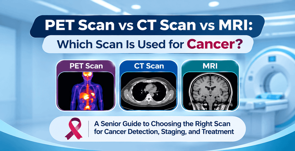
PET Scan vs CT Scan vs MRI: Which Scan Is Used for Cancer?
When facing a potential or confirmed cancer diagnosis, navigating the medical testing process can feel overwhelming. Among the most critical tools in modern oncology are advanced diagnostic imaging scans. Patients and their families frequently ask: What is the difference between a PET scan, a CT scan, and an MRI? Which scan is best for detecting cancer, and why does a doctor order one over another?
Each imaging technology plays a unique, specialized role in cancer detection, staging, treatment planning, and monitoring. While a Computed Tomography (CT) scan and Magnetic Resonance Imaging (MRI) provide detailed structural pictures of organs and tissues, a Positron Emission Tomography (PET) scan detects metabolic activity at the cellular level. In modern oncology, these technologies are often combined such as in a PET CT scan in Bangalore to deliver unmatched diagnostic precision.
If you are looking for a high-precision PET scan in Bangalore or seeking clarity on the right diagnostic pathway, this comprehensive guide breaks down how CT, MRI, and PET scans work, their specific roles in cancer care, key differences, safety considerations, and pricing expectations.
Understanding Cancer Imaging: Why Diagnostic Scans Matter
Cancer diagnostic imaging serves as the bedrock of modern oncology. Without precise imaging, oncologists cannot accurately locate tumors, determine their stage, or evaluate whether a treatment plan is successfully working. Diagnostic scans allow medical teams for:
- Early Detection & Screening: Detect suspicious masses early, often before physical symptoms manifest.
- Accurate Staging: Determine the exact size, location, and spread (metastasis) of malignant tumors.
- Monitoring Therapeutic Response: Assess whether chemotherapy, immunotherapy, Radiation therapy or targeted therapy is shrinking tumors or reducing metabolic activity.
- Recurrence Surveillance: Identify recurrent cancer cells early during post-treatment follow-ups.
Choosing the appropriate scan depends on the suspected cancer type, its location in the body, and whether the physician needs structural details or cellular activity data.
What Is a CT Scan and How Does It Help in Cancer Detection?
A Computed Tomography (CT) scan is a specialized diagnostic imaging procedure that utilizes rotating X-ray equipment and advanced computer algorithms to create detailed cross-sectional images (slices) of bones, blood vessels, and soft tissues inside the body.
How a CT Scan Works in Oncology
During a CT scan, the patient lies on a motorized table that slides through a doughnut-shaped scanner. As the X-ray tube rotates around the patient, it takes multiple cross-sectional images. A contrast dye (often iodine-based) may be administered orally or intravenously to highlight blood vessels, lymph nodes, and soft tissue structures.
When Doctors Recommend a CT Scan for Cancer
CT scans are widely used across all stages of cancer management due to their speed and exceptional structural resolution. Key applications include:
- Primary Tumor Detection: Detecting tumors in the lungs, liver, pancreas, kidneys, and abdominal cavity.
- Staging & Extent: Measuring tumor dimensions and checking whether cancer has invaded nearby blood vessels , lymph nodes or any other organs.
- Biopsy Guidance: Providing real-time visual guidance for needle biopsies to collect tissue samples safely.
- Radiation Planning: Mapping out exact anatomical targets for external beam radiation therapy.
What Is an MRI Scan and When Is It Used for Cancer?
Magnetic Resonance Imaging (MRI) is a non-invasive imaging modality that uses powerful magnetic fields, radio waves, and computer technology to produce highly detailed, high-contrast images of soft tissues, organs, and neurological structures without using ionizing radiation.
How an MRI Scan Works
The MRI scanner creates a strong magnetic field around the patient, aligning hydrogen protons in the body's water molecules. Radiofrequency pulses disturb this alignment. As protons realign, they emit radio signals translated into detailed 2D and 3D anatomical images. A contrast agent containing gadolinium is frequently used to enhance tumor visibility.
Specific Cancer Applications for MRI
MRI excels at differentiating between healthy soft tissue and pathological tumor tissue, making it the preferred imaging choice for:
- Neurological Cancers: Brain and spinal cord tumors, where soft tissue resolution is critical.
- Musculoskeletal Malignancies: Soft tissue sarcomas, bone cancers, and deep joint tumors.
- Pelvic Organ Cancers: Evaluating prostate cancer, cervical cancer, uterine cancer, and rectal cancer staging within the pelvic cavity.
- Breast Cancer Screening: Dense breast tissue evaluation, particularly in high-risk patients or when mammograms are inconclusive.
What Is a PET Scan and Why Is It the Gold Standard in Oncology?
A Positron Emission Tomography (PET) scan is a functional, molecular imaging technique that reveals tissue and organ metabolic activity. Unlike CT and MRI, which map physical anatomy, a PET scan may detect chemical changes at the cellular level before structural changes become visible on traditional scans.
The Science Behind PET CT Imaging
Cancer cells rapidly multiply and exhibit an abnormally high rate of glucose metabolism compared to normal cells. Prior to an oncology PET CT scan Bangalore, a tiny, safe amount of a radiotracer most commonly Fluorodeoxyglucose is injected into the patient's bloodstream.
Malignant cells absorb large amounts of the radiotracer due to higher blood supply and higher metabolic activity. As the radiotracer decays, it emits particles that are detected by the PET scanner. Areas of high metabolic activity illuminate as intense "hot spots" on the image.
Why PET CT is Essential for Cancer Care
Modern facilities utilize integrated PET-CT scanners, which perform both a PET scan and a CT scan in a single session. Combining molecular PET data with structural CT anatomy allows oncologists to:
- Differentiate Benign vs. Malignant: Distinguish between benign Lesion and active cancer cells.
- Accurate Staging: Detects exact location and extent of disease locally and also in distant organs that may appear normal on regular CT scans.
- Early Treatment Response: Evaluates chemotherapy or immunotherapy giving the desired results.
- Recurrence Detection: Detects cancer recurrence at the earliest stage.
PET Scan vs CT Scan vs MRI: Key Differences Explained
Understanding how these three advanced modalities compare side-by-side helps patients comprehend why their oncology team selects specific imaging pathways.
Feature / Aspect | CT Scan | MRI Scan | PET-CT Scan |
Primary Focus | Anatomical structure & bone detail | Soft tissue contrast & detailed anatomy | Metabolic activity combined with structure & extent |
Technology Used | Rotating X-rays | Magnetic fields & radio waves | Radioactive tracer + X-rays |
Radiation Exposure | Moderate (ionizing radiation) | Zero radiation | Low-to-moderate radiotracer + low-dose CT |
Scan Duration | 5 to 15 minutes | 30 to 60 minutes | 2 to 3 hours (includes tracer uptake time) |
Best For | Chest, abdomen, bone trauma, quick screening | Brain, spine, pelvic organs, soft tissues | Full-body cancer staging, metabolic tracking |
Contrast Agent | Iodine-based contrast | Gadolinium-based contrast | Radiotracer (FDG) + IV Iodine CT dye |
Patient Comfort | Open, quiet, fast scanner | Enclosed tube, loud tapping noise | Open ring scanner, quiet scanning phase |
Which Scan Is Used for Specific Types of Cancer?
Oncologists tailor diagnostic imaging selections based on organ biology, cancer behavior, and clinical presentation. Here is a breakdown of preferred modalities by cancer type:
- Lung Cancer: A PET CT scan in Bangalore is the Imaging modality of choice for initial staging. CT scans of the chest are used for initial screening or to determine local extent. .
- Breast Cancer: Mammograms and breast ultrasound provide initial screening, followed by high-resolution MRI for assessing local tissue extent. Full-body PET-CT scans are utilized for staging especially for advanced stages.
- Prostate Cancer: MRI of the pelvis is the gold standard for localized tumor staging and assessing the extent. PSMA PET-CT scans offer unmatched accuracy for detecting recurrent or metastatic disease.
- Lymphoma: PET-CT is the gold standard for staging Hodgkin and Non-Hodgkin Lymphoma, evaluating bone marrow involvement, and confirming complete metabolic remission after chemotherapy.
- Gastrointestinal Cancers may help (Colon, Stomach, Liver, Pancreas): Contrast-enhanced CT scans of the abdomen and pelvis may help in evaluating primary lesions, while PET-CT scans help determine the local extent and distant metastases.A specialised form of PET-CT called a FAPI PET CT may be used in some GI cancers.
- Brain Tumors: Brain MRI with contrast is essential due to high baseline brain glucose uptake, which makes standard FDG PET scans less optimal for brain tissue.
PET Scan Cost in Bangalore & Factors Influencing Pricing
When patients are advised to undergo molecular imaging, understanding the PET scan cost in Bangalore is vital for financial planning. On average, the cost of a full-body PET CT scan in Bangalore starts from ₹15,000 in established cancer care centers.
Key Factors Affecting PET Scan Pricing
- Type of Radiotracer: Standard FDG PET-CT scans are priced lower than specialized radiotracers such as PSMA PET-CT(for prostate cancer) or DOTANOC PET-CT (for neuroendocrine tumors) or FDG PET-CT(for GI cancers), which require specialized Radiotracers.
- Scanner Technology: Advanced digital PET-CT scanners deliver lower radiation doses and higher spatial resolution, slightly influencing procedural pricing.
- Scope of Scanning: Specialized cardiac, brain, or localized organ PET scans may differ in cost compared to standard whole-body scans.
- Hospital Infrastructure & Expertise: Accredited oncology hospitals that provide integrated subspecialty nuclear medicine consultations and rapid report turnarounds ensure higher diagnostic quality.
Financial Guidance at ARC Cancer Hospital
At ARC Cancer Hospital, we prioritize pricing transparency and diagnostic excellence. Our advanced diagnostic imaging wing delivers affordable, high-precision PET CT scans supported by expert nuclear medicine specialists. Contact our helpline to inquire about customized diagnostic packages and insurance coverage.
How to Prepare for Your PET CT Scan in Bangalore
Proper patient preparation is essential for accurate PET CT scan results. Because the radiotracer relies on glucose uptake, non-compliance with preparation rules can distort diagnostic images.
- Strict Fasting Protocol: Fast for at least 6 to 8 hours prior to your scheduled appointment. Only plain water is permitted.
- Blood Sugar Control: Patients with diabetes must inform the scheduling team. Blood glucose levels should ideally be under 150–200 mg/dL prior to tracer injection. Insulin administration must be strictly managed.
- Attire & Metal Removal: Wear warm, comfortable, loose clothing without metal zippers, snaps, or buttons. Leave jewelry and valuables at home.
- Time Allocation: Plan to remain at the center for 2 to 3 hours. After radiotracer injection, you will rest quietly in a designated room for 45 to 60 minutes to allow tracer distribution before scanning.
Choosing the Right PET Scan Centres in Bangalore
Selecting a reliable imaging center impacts diagnostic accuracy, treatment planning speed, and patient comfort. If you are searching for trusted PET scan centres in Bangalore or a specialized cancer diagnostic centre Bangalore, consider these vital criteria:
- Accreditation & Standards: Ensure the diagnostic wing holds NABL and NABH certifications, adhering strictly to AERB safety guidelines.
- Advanced Technology: Modern digital PET-CT technology offers faster acquisition times, reduced scan durations, and significantly lower radiation exposure.
- Expert Nuclear Medicine Team: Accurate interpretation requires experienced, board-certified nuclear medicine physicians working directly alongside surgical, medical, and radiation oncologists.
- Integrated Oncology Care: Choose a comprehensive cancer center where diagnostic findings immediately transition into multidisciplinary tumor board reviews and tailored treatment plans.
Frequently Asked Questions (FAQs)
Q1: Is a PET scan better than a CT scan or MRI for detecting cancer?
Ans: A PET scan is not inherently "better," but rather different. While CT scans and MRIs show anatomical shapes and structural anomalies, a PET scan detects cellular metabolic activity. PET scans often detect cancer earlier at a molecular level, but oncologists usually combine PET with CT (PET-CT) for optimal diagnostic accuracy.
Q2: How much does a PET scan cost in Bangalore?
Ans: The PET scan cost in Bangalore typically starts from ₹15,000. Pricing varies depending on the radiotracer used (e.g., FDG vs. PSMA), scanner technology, and the specific facility.
Q3: Can a CT scan or MRI replace a PET scan in cancer staging?
Ans: No but in some early-stage localized cancers, CT or MRI alone may suffice. However, for comprehensive full-body staging, or evaluating early chemotherapy response, a PET-CT scan provides metabolic insights that CT or MRI cannot replace.
Q4: Is radiation from a PET CT scan safe?
Ans: Yes, PET CT scans operate under strict AERB safety guidelines. The radioactive tracer decays rapidly and leaves the body within hours. The benefits of accurate cancer staging far outweigh the minimal radiation exposure.
Q5: How long does a PET CT scan procedure take?
Ans: The overall appointment takes about 2 to 3 hours. This includes 45 to 60 minutes for radiotracer uptake while resting quietly in a private room, followed by the actual scanning phase which lasts only 15 to 30 minutes.
Q6: Why do I need to fast before a FDG PET scan?
Ans: Fasting for 6 to 8 hours for a FDG PET Scan is essential because the radiotracer behaves like glucose. If elevated blood sugar or insulin is present, normal cells absorb the tracer, obscuring malignant tumor visibility.
Q7: Can diabetic patients undergo a PET CT scan in Bangalore?
Ans: Yes, diabetic patients can safely undergo PET CT scans. However, blood sugar levels must be carefully regulated below 150–200 mg/dL before tracer administration. Our clinical team provides customized diabetic prep protocols.
Q8: Which scan is best for brain cancer detection?
Ans: Brain MRI with contrast is the gold standard for brain tumors because of its superior soft-tissue resolution. Standard FDG PET scans are less effective in the brain due to the organ's naturally high background glucose consumption.
Q9: What is the difference between a PET scan and a PET-CT scan?
Ans: A standalone PET scan shows metabolic activity but lacks detailed structured outlines. A PET-CT scan combines PET metabolic data with structural CT scans simultaneously, allowing doctors to pin metabolic "hot spots" precisely onto anatomical structures.
Q10: Does health insurance cover PET CT scans in Bangalore?
Ans: Most comprehensive health insurance policies cover diagnostic PET-CT scans when prescribed by an oncologist for cancer diagnosis, staging, or treatment evaluation. ARC Cancer Hospital provides cashless insurance TPA support.
Q11: How soon will I get my PET CT scan results?
Ans: At specialized facilities like ARC Cancer Hospital, official PET-CT diagnostic reports reviewed by senior nuclear medicine radiologists are typically available within 24 to 48 hours.
Q12: Is a PET scan painful?
Ans: No, the scan is entirely painless. The only minor discomfort is a small needle prick for the IV tracer injection. You rest comfortably during the uptake and scanning phases.
Q13: What should I do after my PET CT scan is completed?
Ans: You can resume normal activities immediately. We advise drinking plenty of water throughout the rest of the day to help flush the tracer out of your system quickly.
Q14: Where can I find a trusted cancer diagnostic centre Bangalore for a PET CT scan near me?
Ans: ARC Cancer Hospital in Wilson Garden, Bangalore offers state-of-the-art PET-CT imaging, rapid reporting, and direct integration with multidisciplinary oncology care teams. Contact our helpline to schedule your scan.
Types of PET-CT Scans Used for Cancer Diagnosis
PET-CT imaging uses specialized radioactive substances called radiotracers to identify biological activity within the body. Different cancers have different biological characteristics, so nuclear medicine specialists select the appropriate PET radiotracer based on the type of cancer, clinical findings, and purpose of the scan.
The commonly used and specialized PET-CT radiotracers include:
1. FDG PET-CT Scan
Fluorodeoxyglucose (FDG) PET-CT is one of the most widely used molecular imaging techniques in cancer diagnosis. It detects increased glucose metabolism, which is commonly observed in many types of cancer cells.
FDG PET-CT is used for staging, assessing treatment response, and detecting suspected recurrence in selected cases of lung cancer, lymphoma, breast cancer, colorectal cancer, and other malignancies.
2. PSMA PET-CT Scan
Prostate-Specific Membrane Antigen (PSMA) PET-CT is a specialized imaging technique used primarily for prostate cancer.
It identifies cells expressing PSMA and helps detect prostate cancer spread to lymph nodes, bones, and other organs. PSMA PET-CT is particularly useful for staging selected higher-risk prostate cancers and evaluating suspected recurrence after treatment.
3. DOTANOC PET-CT Scan
DOTANOC PET-CT is a specialized molecular imaging scan primarily used for evaluating neuroendocrine tumours.
It targets somatostatin receptors commonly expressed on certain neuroendocrine tumour cells. DOTANOC PET-CT helps identify the primary tumour, evaluate disease spread, assess suspected recurrence, and determine suitability for certain targeted radionuclide therapies.
4. FAPI PET-CT Scan
Fibroblast Activation Protein Inhibitor (FAPI) PET-CT is an emerging molecular imaging technique that targets fibroblast activation protein expressed by cancer-associated fibroblasts in the tumour environment.
FAPI PET-CT is being investigated for its potential role in detecting and evaluating various malignancies, including certain gastrointestinal, and other solid tumours. Its clinical application continues to evolve, and it is not yet a routine replacement for established PET-CT techniques.
5. FDOPA PET-CT Scan
Fluorodopa (FDOPA) PET-CT is a specialized imaging technique that evaluates amino acid transport and related metabolic processes in selected tissues.
It is used in evaluating certain neuroendocrine tumours, medullary thyroid cancer, pheochromocytoma, and selected brain tumours. FDOPA PET-CT may provide valuable diagnostic information when conventional imaging or other PET radiotracers are less informative.
How Is the Right PET-CT Radiotracer Selected?
Not all PET-CT scans use the same radiotracer, and no single tracer is suitable for every type of cancer. The selection depends on the suspected malignancy, tumour biology, previous imaging findings, and whether the scan is intended for diagnosis, staging, treatment response assessment, or recurrence detection.
At ARC Cancer Hospital, the appropriate PET-CT imaging approach can be determined through clinical evaluation and consultation with the treating oncology and nuclear medicine teams.
Conclusion: Making Informed Diagnostic Decisions at ARC Cancer Hospital
When it comes to cancer diagnosis, PET, CT, and MRI scans are complementary tools rather than competing technologies. While CT scans offer exceptional anatomical structural imaging and MRIs excel at soft tissue detail, PET scans reveal metabolic cancer activity at the cellular level. Combined modalities like PET-CT represent the height of diagnostic accuracy in modern oncology.
Choosing the right diagnostic pathway ensures accurate staging, eliminates guesswork, and enables your medical team to design the most effective personalized treatment plan. If you or a loved one require an oncology PET CT scan Bangalore or seek guidance on diagnostic options, ARC Cancer Hospital provides world-class nuclear medicine infrastructure, transparent pricing, and compassionate patient care.
Take the first step toward clear diagnostic answers. Visit ARC Cancer Hospital in Bangalore or book your PET CT scan consultation today.
Need Cancer Care Guidance or a Second Opinion?
Consult senior surgical, medical, and radiation oncology specialists at ARC Cancer Hospital, Wilson Garden, Bangalore.